English English
 |


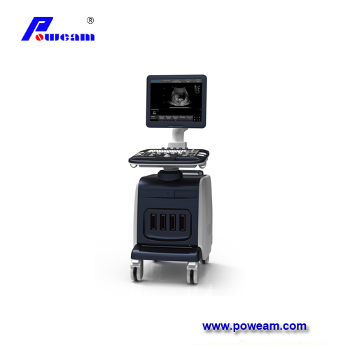
C20E
POWEAM
Color mode:CDFI, PDI (Power Doppler), DPDI (Directional Power Doppler), HR‑Flow;支持 ROI steer,color map V0‑V10、VV0‑VV6、P0‑P3、dP0‑dP3
Spectral Doppler:PW, CW, HPRF;Auto‑calc Doppler hemodynamic parameters(RI,PI,S/D,VTI etc.)
Tissue Doppler:TDI‑TVI / TVD / TVM / TEI (for phased‑array cardiac probe)
Workflow:Dual‑Live B+C mode,Color Panoramic,Raw‑data post‑processing for color doppler images
| No. | Key Specification Point |
|---|---|
| 1 | C20E is a portable ultrasound machine with color doppler flow imaging, supporting multi‑clinical applications including abdomen, obstetrics, gynecology, urology, cardiology, small parts, blood vessel, superficial, orthopedic, MSK, nerve and pediatrics exams. |
| 2 | Equipped with multiple transducer selections: convex, micro‑convex, linear array, phased array, trans‑vaginal and 4D volume probes, compatible with CDFI, PDI, DPDI, HR‑Flow for vascular, obstetrics, gynecology and cardiology examinations. |
| 3 | Rich imaging modes cover B‑Mode, 2B/4B‑Mode, M‑Mode, B/M‑Mode, plus spectral PW CW doppler, CDFI, PDI, DPDI, HR‑Flow, TDI(TVI/TVD/TVM/TEI), Panoramic, Elastic Imaging and optional 3D/4D mode. |
| 4 | Built‑in tissue doppler imaging TDI, dual live B‑color doppler mode and color panoramic doppler ultrasound function; supports trapezoidal, EFOV extended field‑of‑view, SCI spatial composite and FCI frequency composite imaging algorithms. |
| 5 | This system integrates high‑sensitivity power doppler DPDI and puncture enhancement; advanced ZClear, THI tissue harmonic imaging, auto‑optimization and auto‑IMT carotid intima‑media thickness measurement are available for daily clinical workflow. |
| 6 | Hardware configuration: 15.6‑inch 1920×1080 high‑resolution monitor, 3 activated transducer ports, 1TB hard‑disk, Linux operating system, approx. 6.45 kg weight without battery, with 14.4V/6500mAh rechargeable battery module. |
| 7 | Measurement library covers comprehensive 2D, M‑mode and Doppler measurement packages, including auto‑NT, smart OB calculation, auto‑follicle measurement and IMT analysis, suitable for small‑parts, superficial, nerve and MSK diagnostic scenarios. |
| 8 | Complete data management solution: supports RAW/DICOM/JPG/BMP/TIFF image, AVI/MP4 video and PDF report export; enables WiFi, Bluetooth and LAN‑to‑PC transmission, DICOM 3.0 standard communication protocol for hospital PACS connection. |
| 9 | Optional feature list: 3D,4D,Panoramic,Elastic Imaging,ZLive and Silhouette; can combine color doppler with elastic imaging for superficial tissue and organ assessment, multiple optional accessories including trolley, biopsy kit, foot‑switch and thermal printer. |
| 10 | Supports optional static 3D&4D volume rendering; can realize multi‑plane MPR, VR volume reconstruction and auto‑rotation display for obstetrics and gynecology volume scan diagnosis. |